How Cardiologists Use Echocardiograms for Heart Health Diagnosis
Cardiology involves diagnosing and treating diseases of the cardiovascular system, and physicians use various diagnostic tools to assess heart health. One standard procedure is an echocardiogram, which provides detailed images of the heart. Here is more information on cardiology, including what an echocardiogram is, how it works, the process involved, and its benefits:
What Is Cardiology?
Cardiology is a branch of medicine that focuses on the heart and the circulatory system, addressing conditions that affect these areas. Cardiologists are specialists who diagnose and treat disorders such as heart failure, coronary artery disease, and heart rhythm problems. They use a range of diagnostic procedures to evaluate heart function and structure. These diagnostic tools enable cardiologists to develop treatment plans for patients, and they are a fundamental part of patient care.
What Is an Echocardiogram?
An echocardiogram is a non-invasive test that uses sound waves to produce live images of the heart, allowing doctors to monitor how the heart and its valves are functioning. This test provides information about the size and shape of the heart, as well as its pumping capacity. A physician may recommend an echocardiogram to investigate signs or symptoms of heart problems, and it can help diagnose and monitor certain heart conditions over time. The information gathered from an echocardiogram helps determine the next steps in a patient’s treatment plan.
How Does It Work?
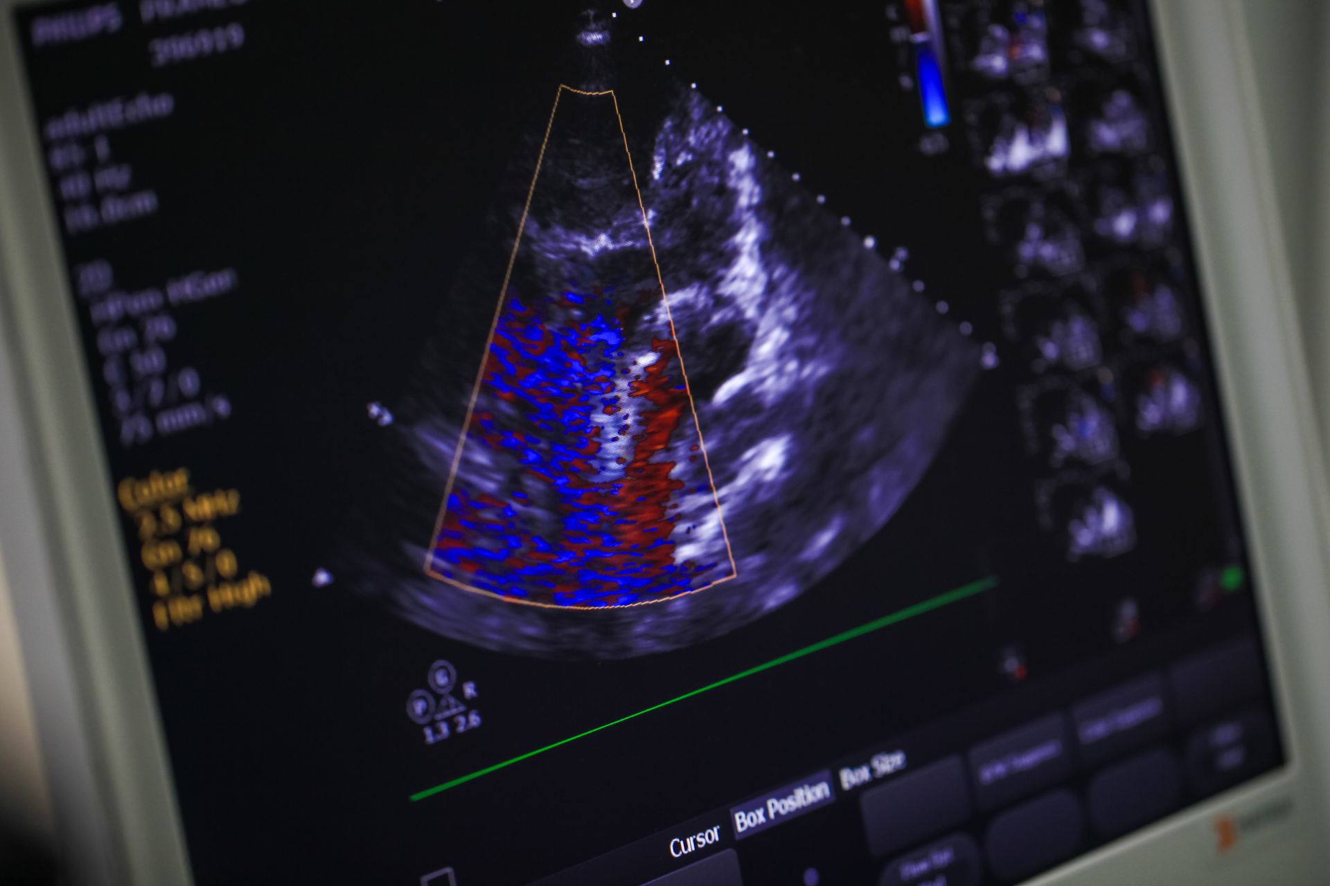
An echocardiogram uses a device called a transducer. This instrument sends high-frequency sound waves through your chest toward your heart. The sound waves bounce, or “echo,” off the heart’s structures, and the transducer picks up the returning waves.
A computer then converts these sound waves into moving images, which are displayed on a monitor, allowing the cardiologist to see the heart beating and pumping blood. Different types of echocardiograms exist, including transthoracic, transesophageal, and stress echocardiograms. Each type offers a distinct perspective on the heart.
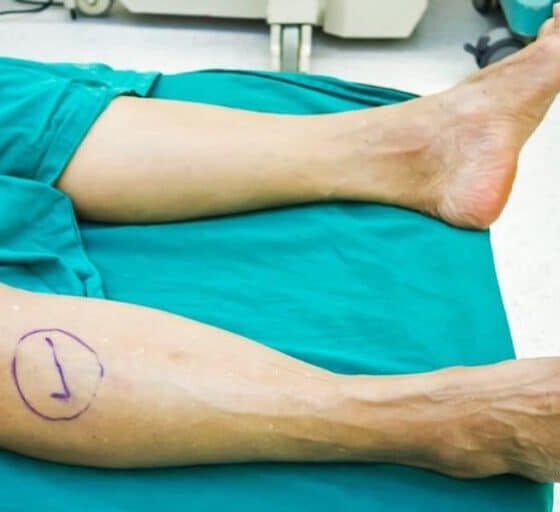
What Does the Process Involve?
During a standard transthoracic echocardiogram, you will lie on an examination table, and a sonographer will apply a special gel to your chest. This gel helps the sound waves travel more effectively, and the sonographer will press the transducer firmly against your skin. The sonographer moves the transducer across your chest to capture different views of your heart, and you may be asked to change positions or hold your breath briefly. The entire procedure is generally painless and typically takes less than an hour to complete.
What Are the Benefits?
An echocardiogram is a valuable diagnostic tool because it offers several benefits for patients and physicians. The procedure is non-invasive, which means it does not require any incisions or instruments to be inserted into the body. This reduces the risks associated with the test.
Echocardiograms do not use radiation, making them a safe option for most people. The test provides real-time images of the heart, allowing for a dynamic assessment of its function and structure. This detailed information aids in the accurate diagnosis and management of heart conditions.
Seek a Heart Health Diagnosis
An echocardiogram is a standard diagnostic test used in cardiology to evaluate the heart’s structure and function, utilizing sound waves to create moving images of the heart. The procedure is non-invasive, safe, and provides detailed information to aid in diagnosing and monitoring heart conditions. Speak with a cardiologist to receive a diagnosis and a suitable treatment plan.
- Family Medicine’s Focus on Mental Health and Wellness
- Understanding the Different Types of Arthritis and Their Impact on Daily Life
- The Benefits of Building a Long-Term Relationship with Your Family Doctor
- Tips for Celebrating Milestone Achievements for Children With Cerebral Palsy
- The Role of Peptide Therapy in Modern Health and Wellness